
Space Spider’s Light Speed Power Has Kids Smiling!

Using 3D scanning technology to make microtia patients whole
Microtia is a condition in which children are born with a malformed ear or no ear at all. Traditional methods of treating this condition have involved numerous lengthy surgeries, which are painful, potentially scarring, and not guaranteed to be successful. Dr. Sheryl Lewin, a craniofacial surgeon, began to look for alternative solutions and was introduced to the Artec Space Spider. She immediately saw the potential for Space Spider’s accurate and lightning-fast hand-held scanning power, especially with children, who tend to have less ability to sit still for a long time.

“I truly love this scanner. I can travel with it so easily because it’s lightweight and easy to carry onto a plane. I’ve brought it to microtia conferences across the United States where I meet new patients and scan their ears in minutes. When they ultimately travel to Los Angeles for surgery, everything’s ready for them.”
This is a quote from Dr. Sheryl Lewin, who has been using Artec Space Spider to create new ears from porous polyethylene for hundreds of children born with microtia. Dr. Lewin deems the Space Spider as the most significant single boost in results throughout her entire career.
Once scanning with the Space Spider begins, full, accurate detail is captured in real time as the structured light technology transforms organic shapes and features into a 3D model in just seconds. This non-invasive technology allows children as young as 4 years old the opportunity to qualify for the outpatient procedure rather than being forced to wait until the age of 10 to even be considered as a candidate for the traditional, painful and potentially scarring surgery.
The Process
Dr. Lewin had already been creating her own sculpted ears as an alternative way to help children with microtia. She felt, though, that her replicas were missing some symmetry that a hand-carved model could not offer. Therefore when she discovered Space Spider, she redesigned the workflow to accommodate this new technique. Now, the workflow looks like this:
Step 1 - Dr. Lewin pre-approves each patient to ensure optimal candidacy for the surgery. She also views the ear carefully, through images and in person, to ensure it is a good candidate for scanning.
Step 2 - the child’s head is prepared for scanning by gelling the hair around the ear up and away from the ear, and a stocking cap is placed over the head to hold the hair in place.
Step 3 - the child is distracted by a computer or cellphone game while Dr. Lewin makes a few passes with the scanner to capture the full ear in high resolution, full color 3D. This imagery is fed back into the Artec Studio software.
Step 4 - Dr. Lewin will then make any last-minute adjustments or refinements to the ear on Artec Studio.
Step 5 - the STL file is then sent to the implant specialist, Poriferous LLC, who makes the new ear out of porous polyethylene. It is shipped within two weeks.
Step 6 - once it has arrived, Dr. Lewing makes some final adjustments so that when it is implanted, it will sit symmetrically on the head, matching the whole ear.
Step 7 - the ear is implanted onto the child’s head.
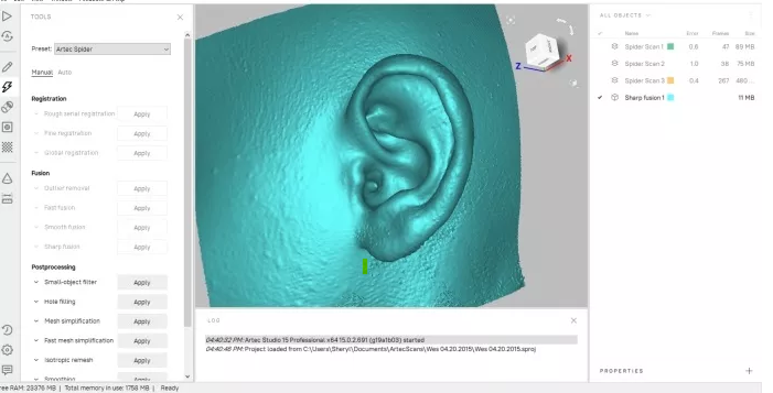
Space Spider scan of a patient’s normal sized ear in Artec Studio software

This is just one more example of how 3D scanning technology is being used in the medical field to improve the knowledge and quality of life of thousands of individuals, young or old, big or small.
If you have a requirement for scanning technology, please contact us today to discuss how we can help you with your project.
The original article can be found here.